44 tem image of chloroplast labeled
(PDF) WESTERN BLOT TECHNIQUE - ResearchGate 01.01.2013 · INTRODUCTION The western blot (sometimes called the protein immunoblot) is a widely accepted analytical technique used to detect specific proteins in … PDF Chloroplasts Structure and Function Factsheet Chloroplasts - Structure and Function Chloroplasts are the site of photosynthesis in green plants. This is the process by which carbon dioxide and water are converted into organic chemicals using light as a source of energy. Oxygen is produced as a very useful by-product of the reaction. This Factsheet explains how the structure of a ...
Chloroplasts - Definition, Structure, Function and Microscopy To view chloroplasts under the microscope, students can use toluidine blue stain to prepare a wet mount. This simply involves the following simple steps: Place a plant sample onto drop of water on a clean glass slide Using a dropper, add a drop of the stain (toluidine blue) on the sample and allow to stand for about a minute

Tem image of chloroplast labeled
PDF Plant Anatomy: Images and diagrams to explain concepts photosynthesis occurs in chloroplasts. Like plant cells, animal cells are also eukaryote cells. Unlike animal cells, plant cells have a cell wall. The cell wall is made of cellulose but may be thickened and strengthened in some cells. PLANT ANATOMY AND PHYSIOLOGY: IMAGES AND DIAGRAMS TO EXPLAIN 5 Figure 1.1. Features of a typical plant cell. Lab #8 photosynthesis with some questions from mito,meio an cyto A: No because a green lights color is not absorbed by plants it is reflected 6) Examine this electron micrograph of a chloroplast a) Identify the stack of membranes labeled A b) Identify the region labeled B c) would the production of organic compounds during light independent reactions occur in region B or on the membranes labeled A Science and medical images, photos, illustrations, video footage ... Permanent Redirect. The document has moved here.
Tem image of chloroplast labeled. Chloroplast: Meaning, Structure, Analogy - Embibe The word chloroplast comes from the Greek words 'khloros', meaning "green", and 'plastes', meaning "formed". The diagram of Chloroplast is given below. Fig: A Labeled Diagram of Chloroplast Chloroplast Structure Chloroplasts are roughly \ (1 - 2\, {\rm {μm}}\) thick and \ (5 - 7\, {\rm {μm}}\) in diameter and are seen in all higher plants. Chloroplast Stock Illustrations - Stock Photos & Royalty Free Photos by ... New users enjoy 60% OFF. 186,942,128 stock photos online. Download 616 Chloroplast Stock Illustrations, Vectors & Clipart for FREE or amazingly low rates! ... An stem epidermis, scanning electron microscopy. Photosynthesis accumulating sugar and cellular respiration fueling all plants functions day night 2 educational posters vector ... Tem And Chloroplasts Stock Photos, Pictures & Royalty-Free Images - iStock Tem And Chloroplasts Stock Photos, Pictures & Royalty-Free Images - iStock Search from Tem And Chloroplasts stock photos, pictures and royalty-free images from iStock. Find high-quality stock photos that you won't find anywhere else. Photos Curated content Curated sets Signature collection Essentials collection Diversity and inclusion sets Chloroplast Photos and Premium High Res Pictures - Getty Images chloroplast structure 4,278 Chloroplast Premium High Res Photos Browse 4,278 chloroplast stock photos and images available, or search for chloroplast micrograph or chloroplast structure to find more great stock photos and pictures. NEXT
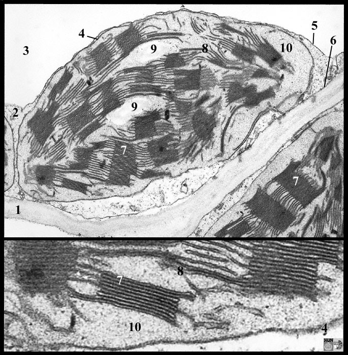
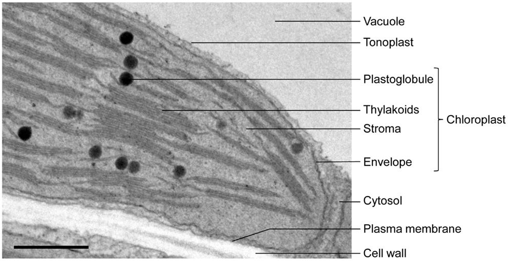
Three-Dimensional Analysis of Chloroplast Structures Associated with ... Chloroplasts are responsible for the eukaryotic photosynthesis and carbon fixation, thus providing energy for much of life on the earth. Chloroplast biogenesis is a complex process and is highly integrated with cellular and plant development (Yang et al., 2010; Pogson et al., 2015).Although three-dimensional (3D) models of the thylakoid membrane architecture have been created using electron ... Structure of Chloroplast (With Diagram) - Biology Discussion This will help you to draw the structure and diagram of chloroplast. 1. Chloroplasts (Figs. 295-296), responsible for the photosynthesis of the plants, are the characteristic features of the cells of green plants. ADVERTISEMENTS: 2. Around the chloroplast is present a double membrane envelope. 3. Each membrane of chloroplasst is 35 to 50 Å thick. Transmission Electron Microscopy (TEM) for Chloroplast? 3) Rinse off excess PFA & GA in 150mM HEPES (pH 7.35) with 30mM MgCl2 3 times for 5 minutes. 4) Place samples in 1% Osmium Tetroxide in 150mM HEPES with 30mM MgCl2 store at 4-degrees for 2-hrs. 5 ... Transmission electron microscopic images of chloroplasts ... - ResearchGate For easy organelle identification, a chloroplast (P) and a mitochondrion (M) are labeled. (C-E) Ultrastructure of chloroplasts and mitochondria in cells of the strong PRORP1 RNAi mutant line...
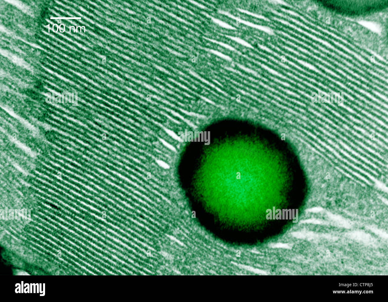
rohrreinigung-notfallservice.de › xawgjpguei › leafLeaf Cell Under Microscope Labeled Jun 19, 2022 · In Figure 2 1, try to make an accurate labeled sketch of the cell showing exactly the detail that your microscope reveals in the best cells from your slides. Make a good microscope drawing of these cells under high power. This is an image of the Osmanthus heterophyllus under a 50x magnification using the boom microscope. Chloroplast High Resolution Stock Photography and Images - Alamy Conceptual image of chloroplast. ID: DNDWWM (RF) The beautiful leaves contain white pigment ID: 2DAM45R (RF) Photosynthesis, illustration ID: 2FYMTR6 (RF) Chloroplast ID: 2GR8A84 (RF) Deficiency of minerals in plant. lack of nitrogen, potassium. Sick yellow currant leaves. ID: 2B1J04J (RF) Chloroplast, plant cell organelle ID: 2G3M4YC (RF) Chloroplasts | Photoreceptor Apparata | Algae - Biocyclopedia Transmission electron microscopy image of a chloroplast at higher magnification showing the thylakoid membrane and the eyespot globules (b). (Bar: 1 µm.) FIGURE 2.79 Transmission electron microscopy image of Nannochloropsis sp. in transverse section, showing the chloroplast (a) (Bar: 0.50 mm); chloroplast at higher magnification (b) (Bar: 0.10 ... Chloroplast- Diagram, Structure and Function Of Chloroplast Diagram of Chloroplast The chloroplast diagram below represents the chloroplast structure mentioning the different parts of the chloroplast. The parts of a chloroplast such as the inner membrane, outer membrane, intermembrane space, thylakoid membrane, stroma and lamella can be clearly marked out.
Basisanatomie
Looking at the Structure of Cells in the Microscope Whereas the TEM uses the electrons that have passed through the specimen to form an image, the SEM uses electrons that are scattered or emitted from the specimen's surface. The specimen to be examined is fixed, dried, and coated with a thin layer of heavy metal. Alternatively, it can be rapidly frozen, and then transferred to a cooled specimen stage for direct examination in the microscope ...
Frontiers | When Proteomics Reveals Unsuspected Roles: The ...
microbenotes.com › under-the-microscopeAmazing 27 Things Under The Microscope With Diagrams May 13, 2022 · Figure: Virus (SARS-CoV-2) under the microscope (TEM). Image Source: NIAID (Flickr). Transmission electron microscope. Transmission electron microscopes are better for the observation of viruses as they provide up to 1000X magnification of particles. Through this type of microscope, it is possible to observe viruses inside the cells of living ...
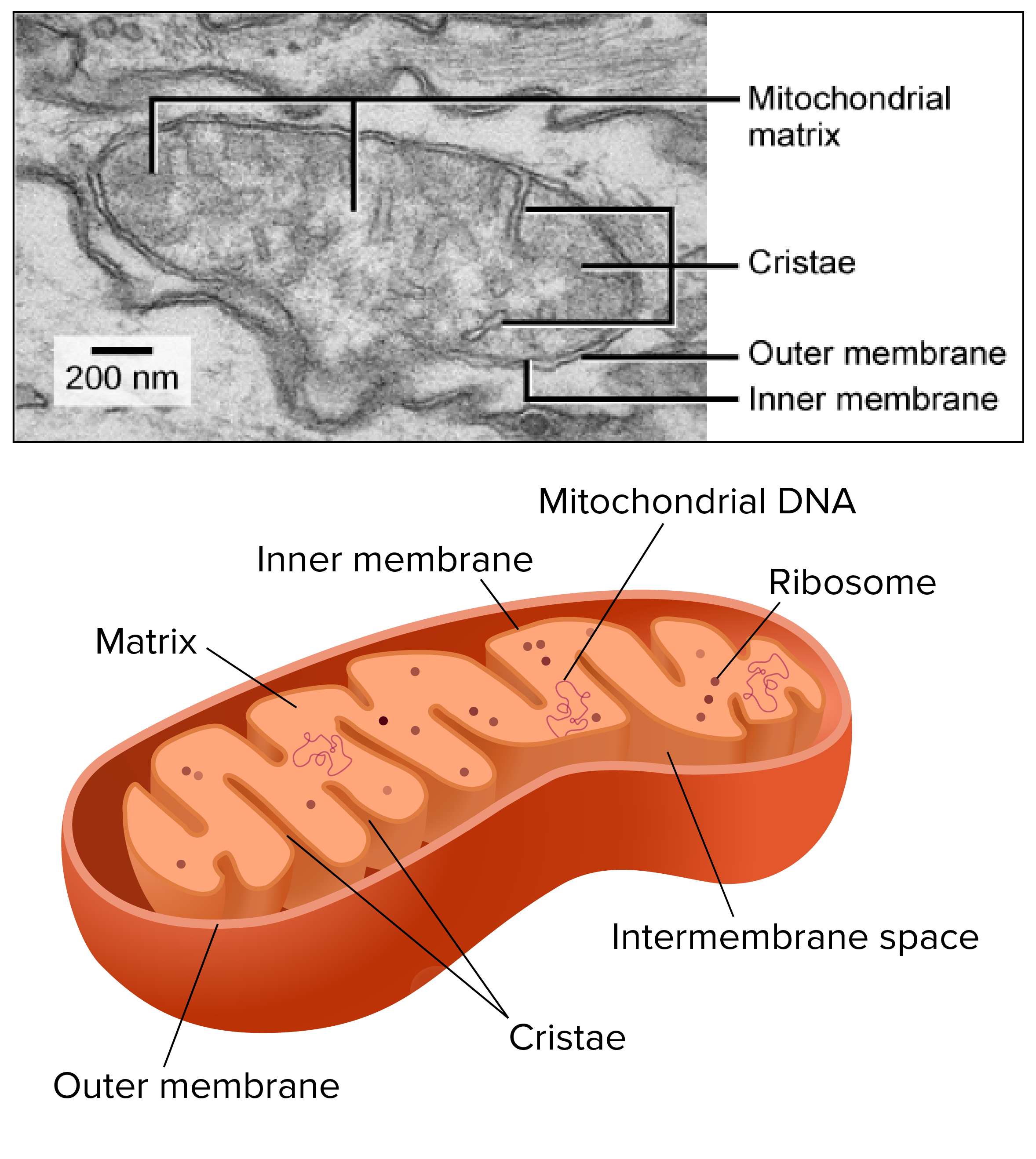
Mitochondria - Membrane Bound Organelles And Defining Characteristics ...
› articles › s41467/022/29961-7Engineering artificial photosynthetic life-forms ... - Nature Apr 26, 2022 · The origin of eukaryotic cells is a fundamental milestone in the evolution of complex life forms, and the evolution of organelles is one of the key steps in eukaryogenesis.

Pin on IB Biology HL
Nanomaterials and nanotechnology for the delivery of … 04.01.2022 · Nanomaterials (NMs) have received considerable attention in the field of agrochemicals due to their special properties, such as small particle size, surface structure, solubility and chemical composition. The application of NMs and nanotechnology in agrochemicals dramatically overcomes the defects of conventional agrochemicals, including low bioavailability, easy photolysis, and organic ...
Plant Cell Chloroplast Stock Photos & Plant Cell Chloroplast Stock ...
› books › NBK26880Looking at the Structure of Cells in the Microscope Confocal microscopy and image deconvolution both provide thin optical sections and can be used to reconstruct three-dimensional images. Determining the detailed structure of the membranes and organelles in cells requires the higher resolution attainable in a transmission electron microscope. Specific macromolecules can be localized with ...

Quia - Cell Organelle Pictures
KMBT 654-20160901143845 - Quia photosynthesis occurs in organelles called chloroplasts. In photosynthesis, carbon dioxide and water are converted into glucose and oxygen. Glucose can be used to make other macromolecules 4. Glucose consists of carbon, oxygen, and hydrogen atoms. 5. Glucose is used as a precursor to make many other

Quia - Section 7.2 Organelle ID (Extra credit opportunity)
PharmaCircle This website uses cookies to help provide you with the best possible online experience. Please read our Terms & Conditions and Privacy Policy for information about ...

Chloroplasts TEM - Stock Image - C008/5120 - Science Photo Library
Ribosome - protein factory - definition, function, structure … Instead, a transmission electron microscope (TEM) is required to view ribosomes and rough endoplasmic reticulum. References “Ribosome” “Ribosome” by British Society for Cell Biology “ Nucleus and ribosomes ” “Structure and Function of the Eukaryotic Ribosome” by Jennifer A Doudna and Virginia L Rath “Ribosome database project”
Post a Comment for "44 tem image of chloroplast labeled"