42 electron micrograph labeled
Endosome - Wikipedia Electron micrograph of endosomes in human HeLa cells. Early endosomes (E - labeled for EGFR, 5 minutes after internalisation, and transferrin), late endosomes/MVBs (M) and lysosomes (L) are visible. Bar, 500 nm. History of cell membrane theory - Wikipedia A more direct investigation of the membrane was made possible through the use of electron microscopy in the late 1950s. After staining with heavy metal labels, Sjöstrand et al. noted two thin dark bands separated by a light region, which they incorrectly interpreted as a single molecular layer of protein. A more accurate interpretation was ...
Here’s why people with allergic asthma are at lower COVID-19 ... May 02, 2022 · Healthy human cells (labeled pink) from the lining of airways grow in “lawns” laced with some mucus (green) in this colorized electron micrograph. C. Ehre, C.B. Morrison et al/PNAS 2022 (CC BY ...
Electron micrograph labeled
Bird Respiratory System - Eastern Kentucky University of blood capillaries, labeled with the presence of erythrocytes (*), and air capillaries (AC) that make up the parabronchi's mantle of gas-exchange tissue (Brown et al. 1997). (A) Micrograph of lung tissue from a Brown Honeyeater ( Lichmera indistincta ) showing (a) parabronchi, (b) blood vessel, and (c) exchange tissue (bar, 200 micrometers). Image Library | CDC Online Newsroom | CDC This electron microscopic (EM) image depicted a monkeypox virion, obtained from a clinical sample associated with the 2003 prairie dog outbreak. It was a thin section image from of a human skin sample. On the left were mature, oval-shaped virus particles, and on the right were the crescents, and spherical particles of immature virions. Phosphatidylinositol-4-phosphate controls autophagosome ... Jul 28, 2022 · Autophagosomes are specialized vesicles that target and deliver cargo to the lytic vacuole. Here the authors show that plasma-membrane derived lipid phosphatidylinositol-4-phosphate supports the ...
Electron micrograph labeled. Structures of L-BC virus and its open particle provide ... Aug 20, 2022 · The electron density map of an empty particle was cropped, normalized, rotated into 222 orientation, the map origin set to the center of the particle, and the crystallographic P23 space group applied. Phosphatidylinositol-4-phosphate controls autophagosome ... Jul 28, 2022 · Autophagosomes are specialized vesicles that target and deliver cargo to the lytic vacuole. Here the authors show that plasma-membrane derived lipid phosphatidylinositol-4-phosphate supports the ... Image Library | CDC Online Newsroom | CDC This electron microscopic (EM) image depicted a monkeypox virion, obtained from a clinical sample associated with the 2003 prairie dog outbreak. It was a thin section image from of a human skin sample. On the left were mature, oval-shaped virus particles, and on the right were the crescents, and spherical particles of immature virions. Bird Respiratory System - Eastern Kentucky University of blood capillaries, labeled with the presence of erythrocytes (*), and air capillaries (AC) that make up the parabronchi's mantle of gas-exchange tissue (Brown et al. 1997). (A) Micrograph of lung tissue from a Brown Honeyeater ( Lichmera indistincta ) showing (a) parabronchi, (b) blood vessel, and (c) exchange tissue (bar, 200 micrometers).
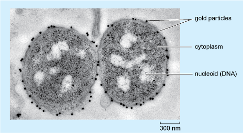
Transmission electron micrograph (TEM) identifying immunogold ...
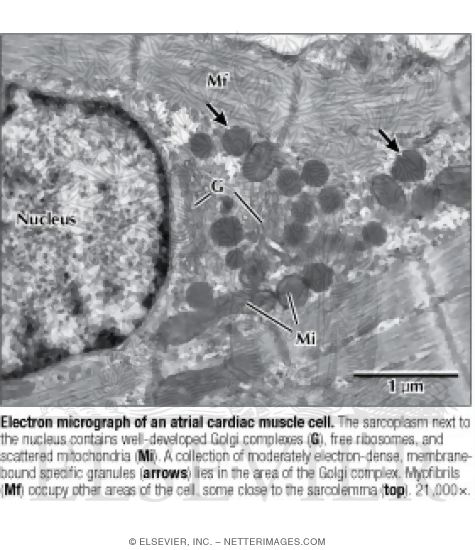
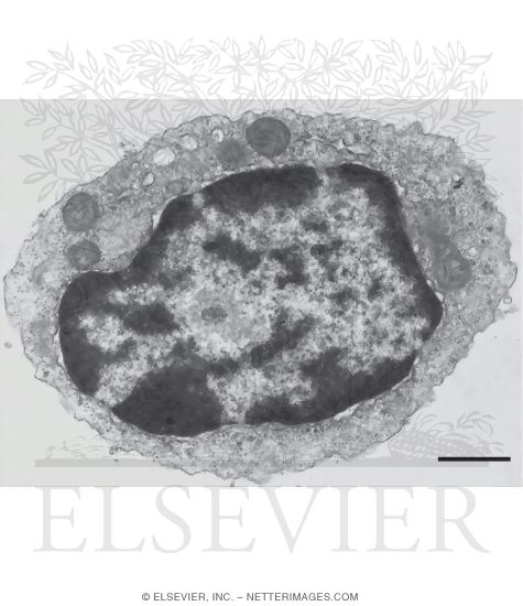
Electron Micrograph of an Atrial Cardiac Muscle Cell

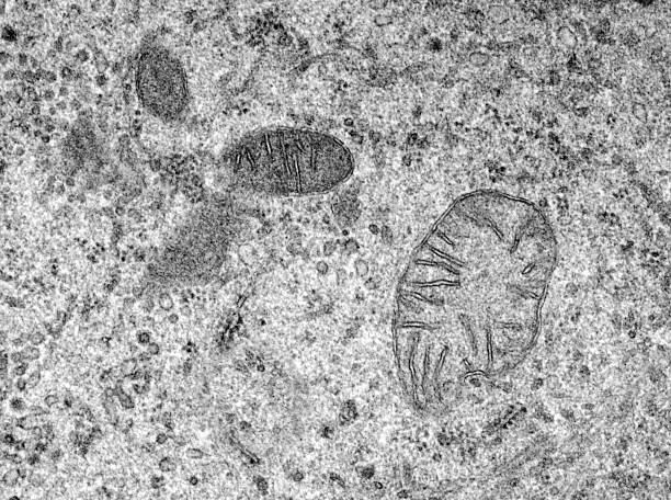
4.9: Eukaryotic Cells - Mitochondria - Biology LibreTexts

1.2 Skill: Interpretation of electron micrographs - YouTube
A tour of the cell: View as single page
Histology Laboratory Manual
Electron Micrographs
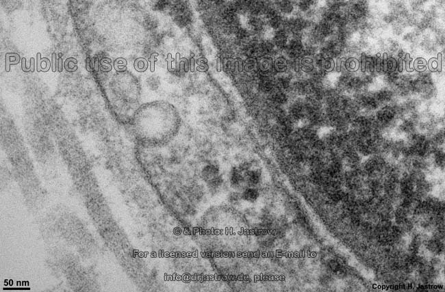
cell and organelles Dr.Jastrow's electron microscopic atlas
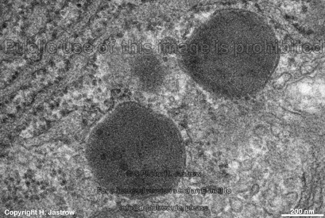
cell and organelles Dr.Jastrow's electron microscopic atlas
7,223 Electron Micrograph Stock Photos, Pictures & Royalty ...
Electron Micrographs

DP Topic 1.1 / 1.2 | Biology - Quizizz
A and B) Electron micrograph of a cell labeled for/5-tubulin ...

Transmission electron micrograph of a thin section through ...

7,223 Electron Micrograph Stock Photos, Pictures & Royalty ...
A tour of the cell: View as single page
cell and organelles Dr.Jastrow's electron microscopic atlas
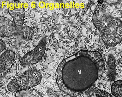
Electron Micrograph of Cell Organelles

Electron micrograph of mouse lung 1 h postinjection of ...
DP Topic 1.1 / 1.2 | Biology - Quizizz
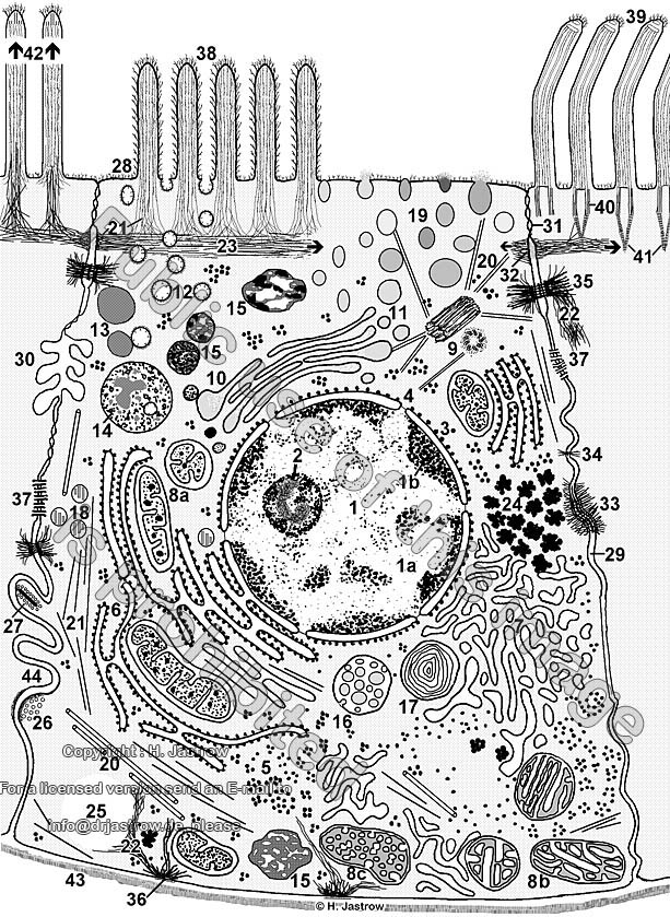
cell and organelles Dr.Jastrow's electron microscopic atlas
IB Biology Notes - 2.3 Eukaryotic cells

Cell Micrographs | BioNinja
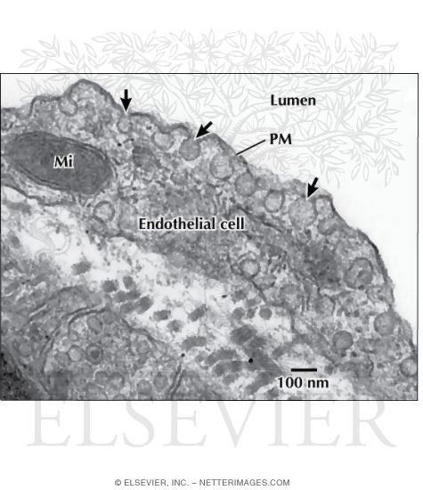
Electron Micrograph of Caveolae and Vesicles In an ...

File:0311 Pancreatic Cells Micrograph labeled.jpg - Wikimedia ...
The Biological bulletin. Biology; Zoology; Biology; Marine ...

Electron micrograph of isolated chloroplasts with the major ...

1. On the following Transmission Electron Micrograph of a ...

DP Biology: Ultrastructure of cells quiz 1.2
Electron Micrographs

Nonmalignant Disorders of Leukocytes Part 1

Electron micrographs of sections through the boundary between ...

AICE Biology Chapter 1: Plant Cell Electron Micrograph ...

Cell Micrographs | BioNinja
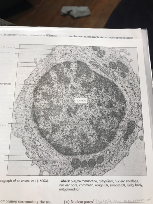
Solved label the ectron micrograph of an animal cell. | Chegg.com
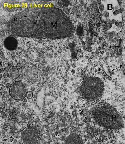
Electron Micrographs
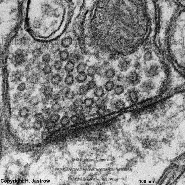
cell and organelles Dr.Jastrow's electron microscopic atlas
A tour of the cell: View as single page
![PDF] Labeling of Hapten-Modified Erythrocytes | Semantic Scholar](https://d3i71xaburhd42.cloudfront.net/da8ea881e5db0ca853c8297ef84adf221a646999/7-Figure6-1.png)
PDF] Labeling of Hapten-Modified Erythrocytes | Semantic Scholar
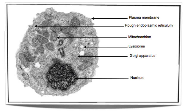
Electron Micrograph of a Lymphocyte

Solved Mitochondrion Nucleus Vesicle Peroxisome | Chegg.com

Electron micrographs of SPIO-labeled MSCs. A, Cell nucleus (N ...
Post a Comment for "42 electron micrograph labeled"