44 how to label gel electrophoresis images
Agarose Gel Electrophoresis for the Separation of DNA Fragments Place an appropriate comb into the gel mold to create the wells. Pour the molten agarose into the gel mold. Allow the agarose to set at room temperature. Remove the comb and place the gel in the gel box. Alternatively, the gel can also be wrapped in plastic wrap and stored at 4 °C until use ( Fig. 1 ). 2. Gel Electrophoresis - an overview | ScienceDirect Topics For gel electrophoresis, a DNA sample is loaded at one end of a gel matrix (usually agarose or acrylamide) that provides a uniform pore size through which the DNA molecules can move. Application of a constant electric field causes DNA fragments (all have a uniform, strong negative charge) to migrate toward the cathode.
GelAnalyzer GelAnalyzer 19.1 Analyze gel images from any source Use your digital camera, smartphone, or gel doc system to obtain images. GelAnalyzer will take care of the rest. Automatic lane and band detection With full manual control over adding, modifying, and deleting lanes and bands. Fix run distortions through Rf calibration

How to label gel electrophoresis images
Polyacrylamide Gel Electrophoresis - Cleaver Scientific The gel mixture is made up not in water but in electrophoresis buffer (Tris-HCl), that provides the ions for electrophoresis. Often, the gel is poured in 2 parts. The first parts is a resolving gel, with a pH around 8.8 which slows the migration of the proteins. Above the resolving gel, a stacking gel is poured with a pH of 6.8 and a larger ... THEV1 - Overview: Thalassemia and Hemoglobinopathy … This evaluation will always include hemoglobins A2 and F and hemoglobin electrophoresis utilizing cation exchange high-performance liquid chromatography (HPLC) and capillary electrophoresis methods. If a serum sample is received, a serum ferritin will always be performed to allow incorporation of possible iron deficiency into profile interpretation and … A Complete Guide for Analysing and Interpreting Gel Electrophoresis Results let see some of the gel images of PCR fragments. 2% gel is required to separate PCR products because PCR products are the smaller fragments of DNA nearly ~100bp to ~1500bp. Image 1: The image is captured under the UV transilluminator instead of the gel doc system to show you the effect of EtBr on the gel electrophoresis results.
How to label gel electrophoresis images. Lysyl Endopeptidase®, Mass Spectrometry Grade (Lys-C) Among the most important techniques in proteome analyses is the in-gel digestion of protein spots/bands that have been resolved by electrophoresis using digestive enzymes, such as trypsin and lysyl endopoptidase. Proteins can be identified by mass spectrometry analysis of the peptides produced by in-gel digestion, and further information regarding post-translational … ImageJ for Editing & Labelling PCR Gel Image - YouTube This Tutorial is all about how to quickly Edit & Label PCR Gel Image Using ImageJ software. Presented by - Elvis SamuelJoin Our Telegram Channel for free Sof... How to Interpret DNA Gel Electrophoresis Results - GoldBio During gel electrophoresis, you may have to load uncut plasmid DNA, digested DNA fragment, PCR product, and probably genomic DNA that you use as a PCR template into the wells. Your digested DNA fragment is a digested PCR product. The next step is to identify those bands to figure out which one to cut. Gel Electrophoresis. Lane 1: DNA Ladder. Gel Electrophoresis - Definition, Purpose and Steps - Biology Dictionary The gel chamber wells are loaded with the DNA samples and usually, a DNA ladder is also loaded as reference for sizes.. 6. Electrophoresis. The negative and positive leads are connected to the chamber and to a power supply where the voltage is set. Turning on the power supply sets up the electric field and the negatively charged DNA samples will start to migrate through the gel and away from ...
Part 2: Analyzing and Interpreting (Agarose) Gel Electrophoresis Results Now let see some real images. The gel image above is the result of restriction digestion. Lane 3, 5, 7, and 8 are a homozygous normal allele with a 184bp band here one band of 68bp is also present, but it is not visible. Lane 2 is a mutant uncut allele of 252bp. Lane 1 and 6 are heterozygous contain three alleles: 252bp, 184bp and 68bp. Addgene: Protocol - How to Run an Agarose Gel Run the gel at 80-150 V until the dye line is approximately 75-80% of the way down the gel. A typical run time is about 1-1.5 hours, depending on the gel concentration and voltage. Note: Black is negative, red is positive. The DNA is negatively charged and will run towards the positive electrode. Always Run to Red. How to make a gel image using Powerpoint - YouTube A quick tutorial on how to make a reasonably polished figure using an image of a gel using Powerpoint. There are certainly more professional ways of doing th... How to quantify each band in gel electrophoresis? - ResearchGate you can do an analytical curve in a 1d gel, with known amounts of bsa for example, use photoshop to quantify the pixels (the curve would be pixels x protein mass you applied for each well) and then...
Gel Electrophoresis: Basics & Steps - SchoolWorkHelper Basic Steps. Aragonese and the buffer are mixed together and microwaved to create the gel. It is poured into a mold and has a "comb" placed in it to make holes for the DNA to be inserted. Once it has cooled the comb is removed. The gel is then placed in the gel electrophoresis box and buffer solution is poured onto it. PV92 PCR - Brian McCauley This is where I'll post the gel images. Video & animation. Alu PV92 Detection by PCR Bio-Rad. We're using the Bio-Rad kit for this lab, so our procedures are more or less the same as shown in this video. How Alu jumps from DNA Learnng Center. Animation explaining how Alu works. Background. Alu elements: know the SINEs. Deininger, 2011. Genome ... Analysis of protein gels (SDS-PAGE) - Rice University Calibrate the gel using standards of known molecular mass (set up a standard curve if necessary) Select polypeptide bands in the lane (s) of interest to be analyzed and identify them by some generic label (e.g., a, b, c,... or 1, 2, 3,...) Estimate molecular mass or relative molecular mass for each band of interest CHAPTER 10 Flashcards | Quizlet Please label the images to review the process of polymerase chain reaction and how its products can be analyzed using gel electrophoresis. Match the components of a typical PCR reaction with the function they serve. ... Please label the images to review the process of screening bacterial clones for those containing a donor gene. Other sets by ...
Genotoxicity Test - Technopreneur International Business Developer Co ...
Solved Please label the images to review the process of - Chegg Question: Please label the images to review the process of polymerase chain reaction and how its products can be analyzed using gel electrophoresis. Dam Deration Denaturation 1 се DNA Replication Pricing Olgorde sha and of of arcon A Cole 770 Restriction andonucleases selectively cleaving sites of DNA cony Piring w Opelweg () Restriction ...

32 How To Label Gel Electrophoresis Images - Best Labeling Ideas
PDF Gel Electrophoresis: How Does It Work - Purdue University 6. Check your gel. If it has cooled enough, it should look opaque (cloudy) now. Check with your teacher if you are unsure if it is ready. If it is ready, move on to step 7 (Caution: follow directions very carefully).

Sanger Sequencing
Annotating A Gel | Get Your Science On Wiki | Fandom Part 1. Photo Editing: 1.Take your JPG or PNG file of your Gel and open it with a photo editing program (GIMP). 2. Under "Image" --> "Transform" rotate your picture by 90 degrees so that your wells are on top of the page. 3. Using the Crop tool Cut out the black borders leaving only the gel. 4.
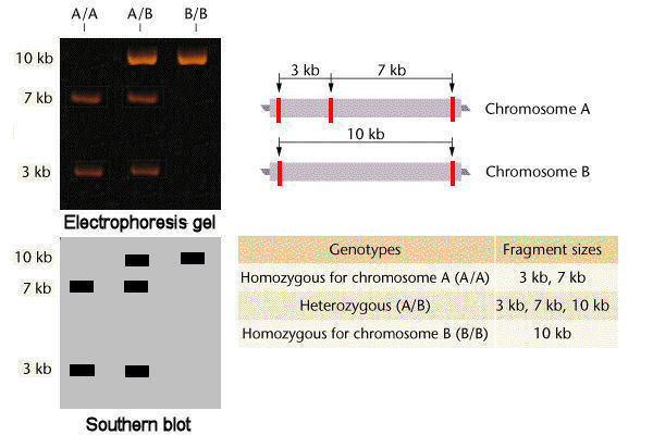
Codominant Molecular Phenotypes
Gel electrophoresis (article) - Khan Academy Gel electrophoresis is a technique used to separate DNA fragments according to their size. DNA samples are loaded into wells (indentations) at one end of a gel, and an electric current is applied to pull them through the gel. DNA fragments are negatively charged, so they move towards the positive electrode.
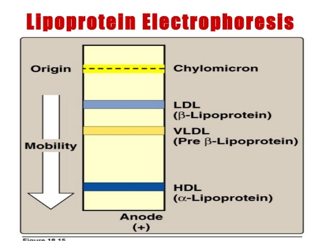
Lecture 3 lipoproteins metabolism-
Solved Please label the images to review the process of - Chegg Science. Biology. Biology questions and answers. Please label the images to review the process of polymerase chain reaction and how its products can be analyzed using gel electrophoresis. Cycle 1 Priming MEGF1-44- MER Sagan 1o Bened d Haut 94C New strand Strands pere SOM 500 5 PERAN Original strands Cydia Amplicon Pos Am Ciprusside parcial ...
Principle of the PCR
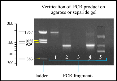
Smearing in agarose gel of PCR product? - ResearchGate 18/05/2022 · Primer dimers (PDs) formed during PCR run is a common finding which be visible after gel electrophoresis of the PCR product. PDs in ethidium bromide-stained gels are typically seen as a 30-50 base ...

Molecular Biology Grade Agarose 500g For DNA Gels - Electrophoresis ...
3 Ways to Read Gel Electrophoresis Bands - wikiHow Hold a UV light up to the gel sheet to reveal results when using a UV-based dye. With your gel sheet in front of you, find the switch on a tube of UV light to turn it on. Hold the UV light 8-16 inches (20-41 cm) away from the gel sheet. Illuminate the DNA samples with the UV light to activate the dye and read the results.
SYPRO™ Ruby Protein Gel Stain
Gel Electrophoresis - University of Utah Gel Electrophoresis. Have you ever wondered how scientists work with tiny molecules that they can't see? Here's your chance to try it yourself! Sort and measure DNA strands by running your own gel electrophoresis experiment. Click to unmute. See how gel electrophoresis is used in forensics. Can DNA Demand a Verdict?

SDS-PAGE - Wikipedia
An overview of technical considerations for Western blotting ... These are required to be from the same blots as the other representative images, and should not be repeated unless the same gel has been stripped and reprobed for multiple targets or different molecular weight targets were measured. As previously discussed, one method of validation of target specificity is the knowledge of a correct molecular weight. Despite this, it is common to …
Post a Comment for "44 how to label gel electrophoresis images"